Abdominal Aortic Aneurysm
What is an abdominal aortic aneurysm?
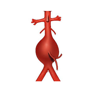
The aorta is our largest blood vessel, delivering oxygenated blood from the heart to the rest of the body. Normally, your aorta’s wall is strong and flexible enough to manage the constant pressure of blood the heart pumps out. The majority of aneurysms are the result of atherosclerosis, a chronic degenerative disease of the artery wall, in which fat, cholesterol, and other substances build up in the walls of arteries and form soft or hard deposits called plaques. If the wall becomes weakened, it can bulge outward forming a balloon-like swelling which can rupture and cause life-threatening bleeding. Aortic aneurysms can occur in the abdomen (abdominal aortic aneurysm/AAA) or in the chest (thoracic aortic aneurysm). Abdominal aortic aneurysms typically develop slowly over a period of many years and hardly ever cause any noticeable symptoms. They are rare in people aged less than 50 years, but the prevalence rises sharply with increasing age.
Why we do this scan
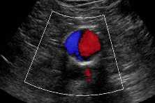
Ultrasound is done to detect aneurysms early, allowing for monitoring and treatment before they rupture. It is a painless, non-invasive test that locates the aneurysm, measures the dimensions and evaluates for plaque build up on the wall which may cause a narrowing.
Why choose us
Ultrasound is an effective and safe tool for the detection of aortic aneurysms and a highly accurate way to measure the size of an aneurysm with experience. Some aneurysms may pose difficulty in viewing due to their location, tortuosity features or calcific plaques limiting penetration of the soundwaves. We have many years of experience navigating these complications to provide your health care provider with accurate measurements and information. We provide your Doctor with extensive anatomical features and if the aneurysm is at a surgical size, further measurements to plan for your best treatment options.
Risk factors
Risk factors include: smoking, having a family member with an abdominal aortic aneurysm, increasing age and being male. AAA's most often affect males, but when they form in females, they have a higher risk of rupturing at smaller sizes as well as occurring in family members. Connective tissue disorders, such as Marfan syndrome and Ehlers-Danlos syndrome and having high blood pressure, cardiovascular disease or high cholesterol are also known risk factors.
Symptoms
Abdominal aortic aneurysm symptoms can include deep, constant belly pain, back or flank pain that may radiate to the legs or buttocks, or a pulsing sensation near the belly button, but many people don't experience symptoms until the aneurysm ruptures, which is a medical emergency.
Signs of a Ruptured AAA (Medical Emergency - seek urgent help):
Severe, sudden abdominal or back pain (may feel like tearing or ripping)
Low blood pressure (dizziness, fainting, shock)
Rapid heartbeat
Cold, clammy skin
Shortness of breath
Loss of consciousness
Complications
If the aneurysm grows too large, it can rupture, leading to life-threatening internal bleeding. Ruptured (burst) abdominal aortic aneurysms are a medical emergency and need immediate treatment with surgery. AAAs larger than 5.0 centimeters in females and 5.5 centimeters in males are particularly at risk and may be considered for surgical intervention. The most common complication is an aneurysm rupture, which causes 150,000 to 200,000 deaths each year around the world.
Preparation for the ultrasond
Fasting (no eating or drinking) is required for 5 hours prior to the ultrasound. Fasting reduces the amount of gas in your abdomen, which provides clearer images of the arteries. All patients should take their usual oral medications with a small amount of water. DO NOT fast if you are a Diabetic. We will ask for you to lie on your back on the examination bed and apply gel to the skin, moving the transducer over the area. The procedure is usually painless and you will hear noises from the ultrasound machine which is the blood flow moving through your arteries.
CoreWave Vascular Ultrasound
Locations:
Suite 3, 46 Buckingham Drive
Wangara, WA 6065
Waikiki Specialist Centre
217 Willmott Drive
Waikiki, WA 6169
Duchess Medical Practice
4/69 Duchess Street
Busselton, WA 6280
More locations to come!
Contact Information:
Email: office@corewave.com.au
Phone: 0437 507 282
Business hours:
8am - 4pm
Monday to Friday
Monthly Saturdays
Fax: (08) 7947 7282