Arms Veins

Arm Veins
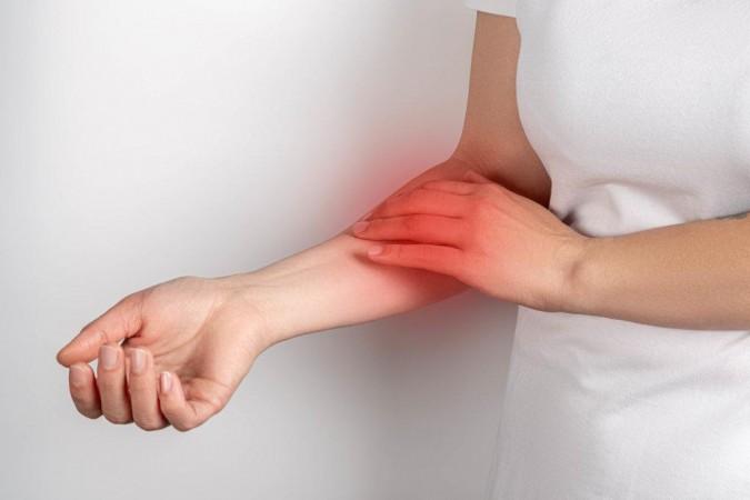
Arm deep vein thrombosis, or upper extremity DVT, is a blood clot that forms in a deep veinous system within the arm. It is less common than DVT in the legs but can still be serious. If left untreated, arm DVT can lead to pulmonary embolism (a potentially life-threatening condition).
Why we do this scan
Ultrasound is the preferred imaging modality for arm DVT due to its non-invasiveness, safety, and accuracy in detecting blood clots. Ultrasound allows for real-time visualization of blood flow, which helps in identifying clots and assessing their impact on circulation. Ultrasound scans may be performed serially over time to monitor the progression of the blood clot and assess the effectiveness of treatment.
Why choose us
Experienced sonographers are preferred for ultrasound imaging of arm deep vein thrombosis due to the complexity of the upper extremity venous system and the need for accurate diagnosis to avoid potentially serious complications. Our skilled sonographer can efficiently identify DVT by assessing vein compressibility, blood flow patterns, and the presence of any thrombi. The upper extremity venous system is more intricate than the lower extremity, with numerous vessels and potential variations that our experienced sonographer can navigate as well as minimize the chances of misdiagnosing a DVT, leading to unnecessary treatment or potentially overlooking a serious condition.
Risk factors
Blood clots in the arm, can be caused by various factors, with central venous catheters (CVCs) and cancer being the most common.
Central Venous Catheters: These are tubes inserted into a large vein, often for medication or fluid administration, and are a major risk factor.
Cancer: Cancer is a significant risk factor, with studies showing a high percentage of cases associated with tumors.
Surgery: Surgical procedures, particularly those involving the upper body, increase the risk of blood clots, including those in the arm.
Injury: Trauma to the arm or surrounding areas can damage veins and increase the risk of clot formation.
Medical Devices: Implants like pacemakers can also contribute to UEDVT.
Other Factors: include smoking, obesity, diabetes, high blood pressure, certain medications (like birth control pills), older age, and a family history of blood clots.
Paget-Schroetter Syndrome: This condition, also known as "effort thrombosis," involves blood clots in the subclavian vein due to repetitive arm movements.
Cervical Rib: A cervical rib can compress the subclavian vein, leading to a blood clot.
Symptoms
Swelling: The arm may swell up, sometimes noticeably larger than the unaffected arm.
Pain: Pain or tenderness may be felt in the arm, often described as cramping or throbbing, especially when standing or walking.
Redness/Skin Color Changes: The skin may appear red, or it may be pale, bluish, or purple in color.
Warmth: The affected arm may feel warm to the touch.
Vein Changes: The veins in the arm may appear swollen, bulging, or more noticeable than usual.
Arm Heaviness: The arm might feel heavy or tired.
Tenderness: The arm may be tender to the touch.
Skin discoloration: The skin may be red or blue.
Complications
This is the most serious complication, occurring when a blood clot from the arm travels to the lungs. A PE can block blood flow to a portion of the lungs, potentially causing tissue death (infarction) and even death. Symptoms include chest pain, shortness of breath, and potentially dizziness or fainting.
Post thrombotic syndrome develops in some individuals after a DVT caused by chronic damage to the veins and valves, with persistent arm pain, swelling, heaviness, and fatigue. Skin changes, such as discoloration, itching, and even ulcers, can also occur due to increased pressure and inflammation in the affected area.
What happens if a DVT is found?
If we find a DVT we will advise you at the time of your scan. We will contact your referring doctor and ask you to wait while we find out what they would like to do. In some cases we may advise you to present to the emergency department of your local hospital. The report findings will be sent to your referring doctor before you leave our rooms or if you are attending the emergency department we will provide you with a printed copy.
Preparation for the ultrasound
There is no patient preparation needed for this scan which will take up to 30 minutes for one arm. You will be asked to remove clothing to expose the neck, shoulder, and arm, however a loose thin strapped singlet may be left on. We will ask for you to either lay down or sit on the edge of our examination bed and the sonographer will apply a gel to the skin and move the transducer over the area. The scan assesses the entire arm from the neck to the wrist, with periodic compressions. The ultrasound examination is generally painless, although some patients may experience discomfort due to probe pressure and we will work to minimise this.


CoreWave Vascular Ultrasound
Locations:
Suite 3, 46 Buckingham Drive
Wangara, WA 6065
Waikiki Specialist Centre
217 Willmott Drive
Waikiki, WA 6169
Duchess Medical Practice
4/69 Duchess Street
Busselton, WA 6280
More locations to come!
Contact Information:
Email: office@corewave.com.au
Phone: 0437 507 282
Business hours:
8am - 4pm
Monday to Friday
Monthly Saturdays
Fax: (08) 7947 7282