Arteriovenous Fistula
What is an Arteriovenous Fistula
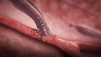
An arteriovenous fistula (AVF) is surgically created by connecting an artery directly to a vein, typically in the arm, to provide a reliable access point for hemodialysis for people with kidney failure. This connection allows for increased blood flow and pressure in the vein, which enlarges and strengthens it, making it suitable for repeated needle insertions during dialysis.
Why we do this scan
Ultrasound is used to create arteriovenous fistulas for hemodialysis access because it allows for precise mapping of blood vessels, ensuring the fistula is created in the best possible location and with optimal vessel size and characteristics. This improves the chances of successful fistula maturation and long-term functionality, reducing the risk of failure and complications. Post-operatively, ultrasound is used to assess fistula maturation, identify potential complications like stenosis or thrombosis, and guide interventions to salvage failing fistulas.
Why choose us
Expertise in ultrasound imaging and interpretation is essential for optimizing patient outcomes in dialysis access. An experienced sonographer is crucial in identifying suitable arteries and veins, evaluating their size and flow characteristics for the successful creation of an arteriovenous fistula. Our sonographer possesses this expertise in interpretation to ensure optimal fistula placement and functionality of the maturing fistula.
Risks
Creating an arteriovenous fistula carries potential risks. These include infection, bleeding, and the formation of blood clots.The fistula or the involved blood vessels may narrow (stenosis) or swell (aneurysm), potentially impacting dialysis or requiring further intervention. Additionally, the fistula can lead to steal syndrome, where the fistula draws too much blood away from the hand, causing pain, numbness, or coldness. More serious complications like heart failure and stroke are also possible, particularly with large fistulas.
Importance of Monitoring
It's crucial to check for a "thrill" or buzzing sensation over the fistula daily, which indicates proper blood flow. Promptly report any signs of infection, bleeding, or other complications to your healthcare provider. Regular monitoring and management of complications are essential for maintaining the health and function of the fistula.
Preparation for the ultrasound
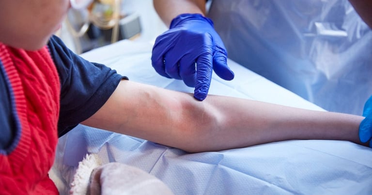
There is no patient preparation needed for this scan which will take up to 30 minutes for each arm. You will be asked to remove clothing to expose the neck, shoulder, and arm, however a loose thin strapped singlet may be left on. We will ask for you to sit on the edge of our examination bed or chair and the sonographer will apply a gel to the skin and move the transducer over the area. The scan assesses the entire arm from the wrist to the neck, with particular attention around the surgical site. The ultrasound examination is generally painless, although some patients may experience mild discomfort due to probe pressure.

CoreWave Vascular Ultrasound
Locations:
Suite 3, 46 Buckingham Drive
Wangara, WA 6065
Waikiki Specialist Centre
217 Willmott Drive
Waikiki, WA 6169
Duchess Medical Practice
4/69 Duchess Street
Busselton, WA 6280
More locations to come!
Contact Information:
Email: office@corewave.com.au
Phone: 0437 507 282
Business hours:
8am - 4pm
Monday to Friday
Monthly Saturdays
Fax: (08) 7947 7282