Carotid Arteries

Carotid Artery Disease
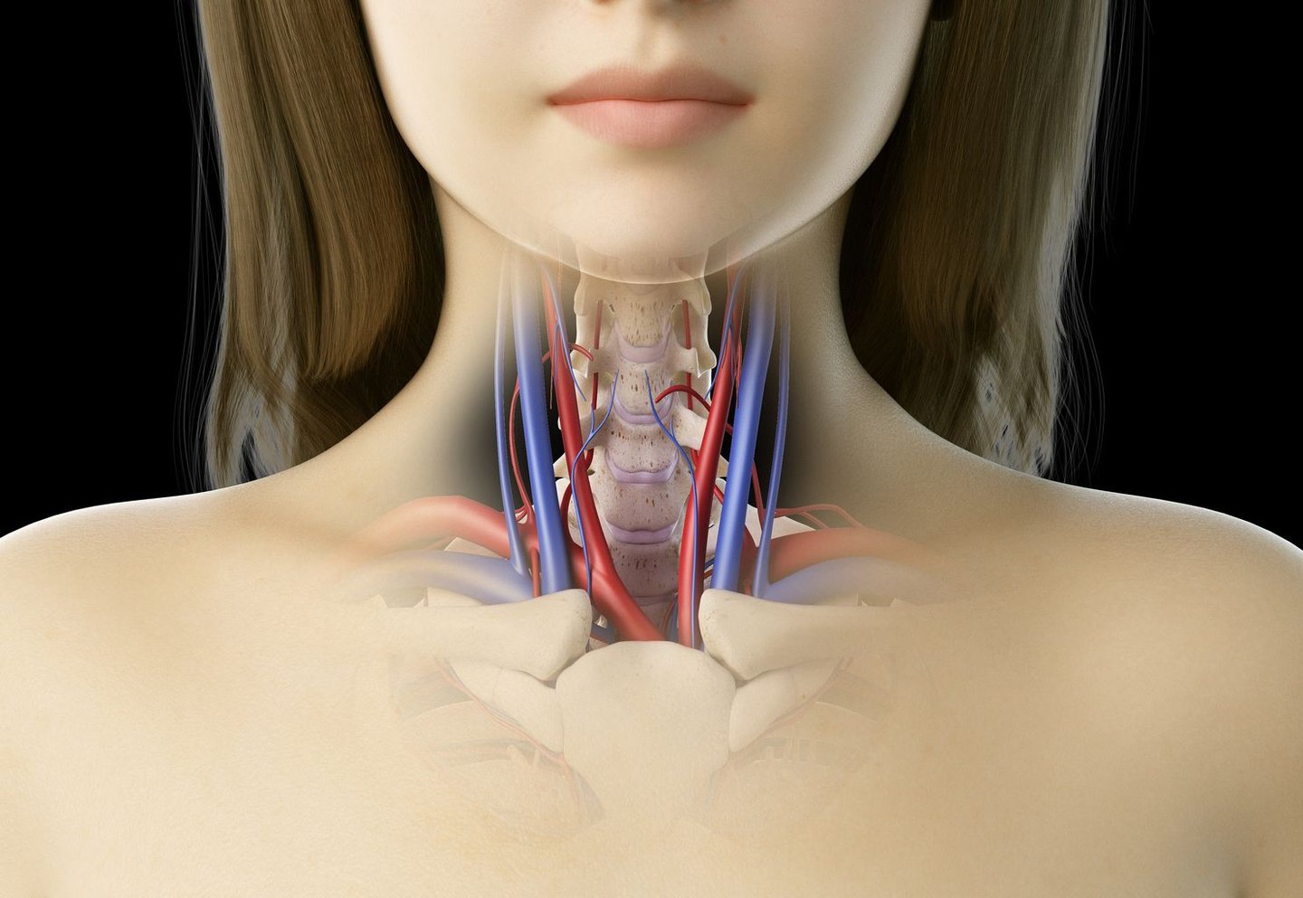
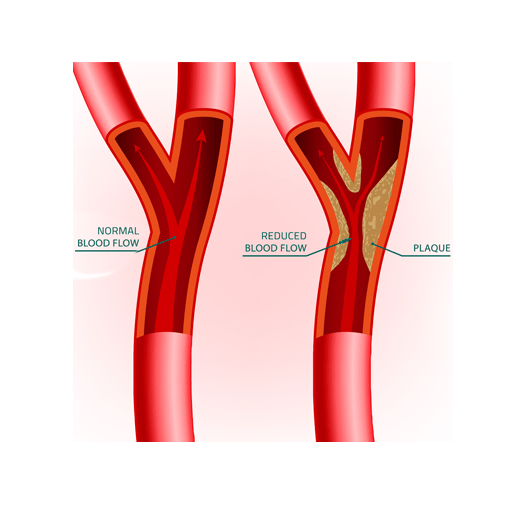
The carotid arteries are major blood vessels that deliver oxygen-rich blood from the heart to the brain and head, with branches supplying the face, neck, and eyes. There is a carotid artery on each side of the neck which divide into two arteries called the internal carotid and external carotd artery. Carotid artery disease occurs when fatty deposits, called athersclerotic plaques, accumulate in the vessel and can lead to narrowing (stenosis) or complete blockage of the artery.
Other rare pathologies are carotid artery aneurysms - a bulge or weakening in the wall of the artery, potentially leading to stroke or other complications if it ruptures or clots. And a carotid body tumor, typically benign (non-cancerous), they can grow and potentially compress nearby nerves and blood vessels.
Why we do this scan
A carotid artery ultrasound, also known as a carotid duplex or Doppler scan, is the principal investigation that assesses blood flow, detecting any blockages or narrowings, between the heart and the brain, that could increase the risk of stroke. It can classify the type of plaque to indicate any plaque instability and increased stroke risk and monitor disease progression.
Why choose us
A standard carotid doppler evaluates the common carotid, internal and external carotid arteries and vertebral artery. Due to the small risk of stroke involving disease of the subclavian and brachiocephalic arteries, we extend our scan beyond the standard protocols to incude and comment on these vessels. We not only assess the common regions for carotid artery disease, but continue along the vessel as far as the ultrasound can attain. And in the case of likely surgical intervention we provide anatomical measurements to assisst in these procedures.
Risk factors
The primary risk factors for carotid artery disease, include high blood pressure, smoking, high cholesterol, diabetes, obesity, lack of physical activity, and a family history of heart disease or stroke. Certain medical conditions, such as kidney disease, autoimmune disorders, and a history of heart disease and peripheral artery disease, increase the risk of carotid artery disease.
Symptoms
Carotid artery disease may not cause any symptoms until a stroke or transient ischaemic attack (TIA) occurs. Symptoms of a stroke or TIA can include sudden numbness or weakness, especially on one side of the body, difficulty speaking or understanding speech, sudden severe headache, loss of vision in one or both eyes, dizziness or loss of balance and difficulty swallowing.
Carotid body tumors, often asymptomatic initially, may present as a painless neck mass and can lead to symptoms like hoarseness, difficulty swallowing, and in some cases, pulsatile tinnitus or high blood pressure.
Complications
The main complication of carotid artery disease is a stroke, where blood flow to the brain is disrupted or blocked. Stroke can cause permanent brain damage, muscle weakness, difficulty speaking, and in severe cases, death. A TIA, also known as a "mini-stroke," occurs when a blood clot briefly blocks a blood vessel to the brain, causing stroke-like symptoms that resolve within 24 hours, usually within an hour. TIAs are a warning sign of a potential stroke and require prompt medical attention to reduce the risk of a future stroke. Since the carotid arteries also supply blood to the eyes, narrowing or blockage can lead to reduced blood flow to the eyes and this can cause blurred vision, sudden vision loss in one eye, or even permanent vision loss.
Preparation for the ultrasond
There is no patient preparation needed for this scan. We will ask for you to lie on your back on the examination bed for the period of the scan which can take up to 20 minutes. A gel is applied to the skin, moving the transducer over the area and you will hear noises from the ultrasound machine which is the blood flow moving through your arteries. Wheelchair bound patients are able to be scanned in the seated position.
CoreWave Vascular Ultrasound
Locations:
Suite 3, 46 Buckingham Drive
Wangara, WA 6065
Waikiki Specialist Centre
217 Willmott Drive
Waikiki, WA 6169
Duchess Medical Practice
4/69 Duchess Street
Busselton, WA 6280
More locations to come!
Contact Information:
Email: office@corewave.com.au
Phone: 0437 507 282
Business hours:
8am - 4pm
Monday to Friday
Monthly Saturdays
Fax: (08) 7947 7282