Deep Vein Thrombus
What is deep vein thrombosis?
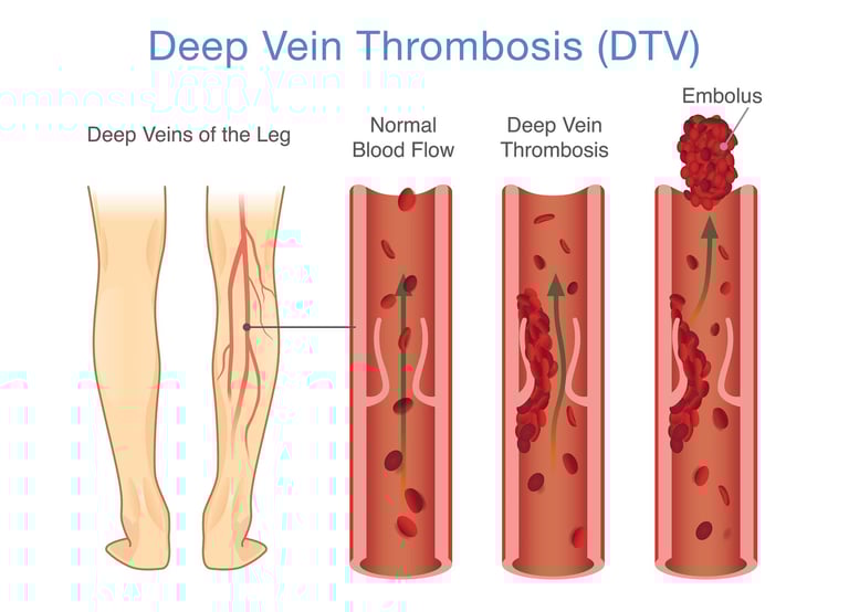
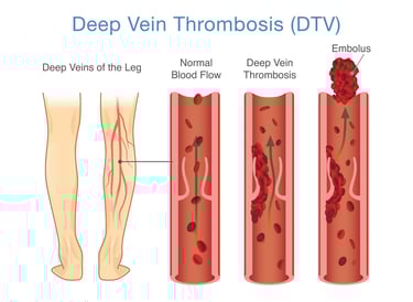
Deep vein thrombosis (DVT) is a condition where a blood clot forms in a deep vein, a major vessel, usually in the legs. If the clot dislodges, it can travel to the lungs and block an artery in the lung, leading to a life-threatening pulmonary embolism. DVT can potentially causing pain, swelling, and warmth in the affected area, but sometimes there are no noticeable symptoms.
Why we do this scan
Ultrasound is the standard imaging test to detect deep vein thrombosis. An ultrasound can determine the size and location of the clot, as well as monitor the effectiveness of treatment. Leg pain and swelling can have various causes, and not all cases are due to DVT. An ultrasound scan helps distinguish DVT from other conditions that may present with similar symptoms, such as muscle strains, cellulitis, or a clot in the superficial veins.
Why choose us
While highly accurate, ultrasound may have challenges visualising clots in the swollen and painful leg or it may be in a challenging location. There are many anatomical variations of the veins which all need to be assessed. Our Sonographer is highly experienced in visualising these veins and their variations through these challenges for accurate assessment.
Risk factors
Factors that increase the risk of DVT include prolonged immobility (like long flights or hospital stays), obesity and a family history of blood clots and certain medical conditions. Cancer, heart disease, lung disease, and certain autoimmune disorders (such as lupus or inflammatory bowel disease) are hypercoagulable states and hormonal changes, such as those during pregnancy, use of oral contraceptives (birth control pills), hormone replacement therapy, or certain fertility treatments, can increase the risk of DVT.
Symptoms
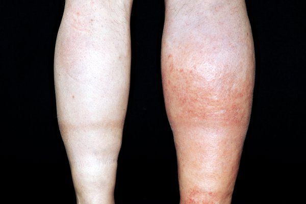
DVT can cause pain, swelling, warmth and redness or discolouration in the skin of the affected leg, but sometimes there are no noticeable symptoms.
Complications
The most serious complication of a DVT is a pulmonary embolism, where a blood clot dislodges and travels to the lungs, potentially causing a blockage and life-threatening consequences. Another long-term complication is post-thrombotic syndrome where damage to the vein valves leads to blood pooling in the legs and increased pressure with chronic swelling, pain, swelling and skin changes.
What happens if a DVT is found?
If we find a DVT we will advise you at the time of your scan. We will contact your referring doctor and ask you to wait while we find out what they would like to do. In some cases we may advise you to present to the emergency department of your local hospital. The report findings will be sent to your referring doctor before you leave our rooms or if you are attending the emergency department we will provide you with a printed copy.
Treatment
Treatment for DVT primarily focuses on preventing the clot from growing and traveling to the lungs, using blood thinners and, in some cases, thrombolytics. If anticoagulants are not an option or not effective, a small filter device may be placed in the inferior vena cava (a major vein in the abdomen) to prevent blood clots from traveling to the lungs. Compression stockings are often recommended to help improve blood flow in the legs and reduce swelling and may need to be worn for several months or longer.
Preparation for the ultrasond
There is no patient preparation needed for this scan which will take up to 30 minutes for each leg. The scan assesses the entire leg from the groin crease to the ankle. We will require you to remove your shoes, socks and trousers/shorts. Very loose shorts, dresses and skirts don't need to be removed and can be lifted out of the way. We will ask for you to lay on your back on the bed with the hip rotated outwards. The procedure is usually painless and involves the sonographer applying a gel to your skin and moving the transducer over the area.
CoreWave Vascular Ultrasound
Locations:
Suite 3, 46 Buckingham Drive
Wangara, WA 6065
Waikiki Specialist Centre
217 Willmott Drive
Waikiki, WA 6169
Duchess Medical Practice
4/69 Duchess Street
Busselton, WA 6280
More locations to come!
Contact Information:
Email: office@corewave.com.au
Phone: 0437 507 282
Business hours:
8am - 4pm
Monday to Friday
Monthly Saturdays
Fax: (08) 7947 7282