Popliteal Entrapment Syndrome


Popliteal Entrapment Syndrome
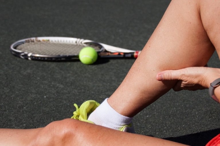
Popliteal artery entrapment syndrome is a rare condition where the popliteal artery, which supplies blood to the lower leg and foot, is compressed by structures behind the knee, usually muscles. This compression restricts blood flow, causing pain and cramping, especially during exercise. The popliteal artery may be positioned abnormally or the calf muscles may be unusually shaped, leading to compression. In some cases, particularly in athletes, the calf muscles may become enlarged due to exercise, putting pressure on the artery.
Why we do this scan
Ultrasound is used to diagnose and assess popliteal artery entrapment syndrome because it can visualize the popliteal artery and its surrounding structures, including the gastrocnemius muscle, and assess blood flow in real-time during different foot and leg positions. This helps identify the type and severity of entrapment and guide treatment decisions.
Why choose us
A high level of skill is crucial for an accurate ultrasound diagnosis of popliteal artrry entrapment syndrome because the condition is complex, involves dynamic compression, and requires specific techniques and interpretation skills. Proficiency is essential as the ultrasound findings help guide treatment decisions, such as whether surgery is needed and the best approach. Our highly skilled vascular sonographer will identify subtle changes in blood flow during various maneuvers to differentiate popliteal artery entrapment syndrome from other conditions.
Risk factors
Popliteal artery entrapment syndrome is more common in young, active males. Risk factors include being a young male (especially in their late teens or 20s), engaging in strenuous athletic activity like running, cycling or weight training, and having certain anatomical variations of the gastrocnemius muscle or popliteal artery. Some individuals are born with an abnormal positioning of the gastrocnemius muscle or popliteal artery, which can predispose them to entrapment. Over time, calf muscles can hypertrophy due to exercise, potentially compressing the popliteal artery.
Symptoms
Pain and cramping in the calf: This is the most common symptom, typically triggered by physical activity and relieved by rest.
Cold or numb feet: Reduced blood flow can make the feet feel cold or numb, especially after exercise.
Tingling or burning sensations: These sensations may be felt in the calf or foot.
Swelling: Swelling in the calf area can occur due to reduced blood flow.
Changes in skin color: The skin on the affected leg or foot may appear pale or bluish, especially after exercise.
Weakness in the leg: Reduced blood flow can lead to muscle weakness.
Complications
Early diagnosis and treatment are important to prevent long-term damage to the artery, which could lead to chronic vascular issues. Untreated cases can cause progressive narrowing of the artery (stenosis), blood clots (thrombosis), and even limb-threatening ischemia or amputation. Chronic compression can lead to the development of a popliteal aneurysm, a bulge in the artery wall and damage to the nerves and muscles in the leg, potentially leading to permanent impairment.
Preparation for the ultrasound
There is no patient preparation needed for this scan which will take up to 30 minutes for each leg. We will require you to remove your shoes, socks and trousers/shorts. Very loose shorts, dresses and skirts don't need to be removed and can be lifted out of the way. We will ask for you to lay in various positions, on your back on the bed with the hip rotated outwards, on your side and in the standing position. The scan assesses the entire leg from the groin crease to the ankle with particular attention behind the knee while the patient performs various leg movements.The procedure is usually painless and involves the sonographer applying a gel to your skin and moving the transducer over the area.

CoreWave Vascular Ultrasound
Locations:
Suite 3, 46 Buckingham Drive
Wangara, WA 6065
Waikiki Specialist Centre
217 Willmott Drive
Waikiki, WA 6169
Duchess Medical Practice
4/69 Duchess Street
Busselton, WA 6280
More locations to come!
Contact Information:
Email: office@corewave.com.au
Phone: 0437 507 282
Business hours:
8am - 4pm
Monday to Friday
Monthly Saturdays
Fax: (08) 7947 7282