Renal Doppler
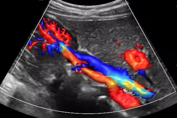
Renal Doppler
A Renal Doppler is an ultrasound routinely performed because it is a useful technique for evaluating blood flow through the renal arteries supplying the kidneys. A narrowing of these arteries is most often caused by atherosclerosis, a build up of plaque (fat and cholesterol) in the artery walls. Another significant cause is fibromuscular dysplasia (FMD), where the artery wall develops abnormally and Takayasu's arteritis where the vessel becomes inflammed.
Why we do this scan
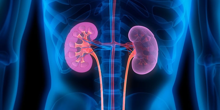
Reduced blood flow to the kidneys can affect their ability to filter waste and excess fluid from the blood, potentially leading to kidney disease. Narrowing of the renal arteries prevents oxygen-rich blood from reaching your kidneys, potentially leading to high blood pressure, kidney damage, fluid retention, electrolyte imbalances, and an increased risk of cardiovascular events. The Doppler ultrasound can also be used to monitor the blood flow to a transplanted kidney.
Why choose us
We have the in-depth understanding of normal anatomy, as well as important anatomical variants, necessary for correct interpretation. The interpretation of renal Doppler examinations is challenging for those with limited experience or those unfamiliar with fundamental concepts and nomenclature. The deep location of both kidneys in the retroperitoneum with relatively small caliber renal arteries further adds to technical challenges associated with this study, and an experienced vascular sonographer is paramount.
Risk factors
Renal artery stenosis is more likely in individuals with certain risk factors like high blood pressure, high cholesterol, diabetes, smoking, and a family history of heart disease. Other factors include advanced age, obesity, and a lack of exercise. An ultrasound might be recommended if you have high blood pressure that is difficult to control, especially if it starts suddenly or worsens without explanation, worsening kidney function, or unexplained kidney shrinkage. Other symptoms might include a whooshing sound (bruit) in the abdomen, fluid retention leading to swelling in the legs and ankles, and even shortness of breath due to fluid buildup in the lungs.
Complications
If left untreated, renal stenosis, can lead to serious complications like high blood pressure, chronic kidney disease, and even kidney failure, potentially requiring dialysis or a kidney transplant.
Preparation for the ultrasond
Fasting (no eating or drinking) is required for 5 hours prior to the ultrasound. Fasting reduces the amount of gas in your abdomen, which provides clearer images of the arteries. All patients should take their usual oral medications with a small amount of water. DO NOT fast if you are a Diabetic. The procedure is usually painless and involves the sonographer applying a gel to your abdomen and moving the transducer over the area. You may be asked to lie down, sit, or stand, with a number of movements such as rolling onto your side and breath holding to optimize the visualization of the kidneys and renal arteries.

CoreWave Vascular Ultrasound
Locations:
Suite 3, 46 Buckingham Drive
Wangara, WA 6065
Waikiki Specialist Centre
217 Willmott Drive
Waikiki, WA 6169
Duchess Medical Practice
4/69 Duchess Street
Busselton, WA 6280
More locations to come!
Contact Information:
Email: office@corewave.com.au
Phone: 0437 507 282
Business hours:
8am - 4pm
Monday to Friday
Monthly Saturdays
Fax: (08) 7947 7282