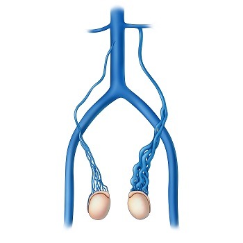
Testicular Veins
What is a varicocele
A varicocele is a common medical condition characterized by abnormal enlargement of the scrotal venous pampiniform plexus, which drains blood from each testicle, resulting in a complex network of swollen vessels. While many individuals with varicoceles may remain asymptomatic, these enlarged veins can lead to various issues, including discomfort, testicular atrophy, and impaired fertility. Varicoceles occur in approximately 15% to 20% of males but are found in about 40% of infertile males. Varicoceles are far more common in the left testicle with a 30% to 40% probability of also occuring on the right.[
Varicocele embolisation
Varicocele embolization is a minimally invasive procedure used to treat varicoceles. It involves using image guidance to insert a catheter into a vein in the groin or neck, and then guiding it through the testicular vein towards the varicocele. Tiny coils or a liquid substance are then used to block the abnormal vein, redirecting blood flow and reducing pressure. This procedure is an alternative to surgery and is typically performed as a day case.
Why we do this scan
The testicular vein ultrasound will map the size and the course of the testicular vein, determining the best treatment approach for the varicocele. Vein mapping ensures that the chosen veins are suitable for the procedure.
Why choose us
Testicular veins can be small and they are located deep in the pelvis, making them challenging to locate and visualize. An experienced vascular sonographer knows the anatomy and can effectively use ultrasound to identify these veins. Our skilful sonographer will provide accurate interpretation of the ultrasound images, crucial for proper diagnosis and treatment and minimising the risk of misdiagnosis to ensure patients receive appropriate care.
Preparation for the ultrasound
Fasting (no eating or drinking) is required for 5 hours prior to the ultrasound. Fasting reduces the amount of gas in your abdomen, which provides clearer images of the arteries. All patients should take their usual oral medications with a small amount of water. DO NOT fast if you are a Diabetic. We will ask for you to lie on your back on the examination bed and apply gel to the skin, moving the transducer over the area. The procedure is usually painless and you will hear noises from the ultrasound machine which is the blood flow moving through your veins.
CoreWave Vascular Ultrasound
Locations:
Suite 3, 46 Buckingham Drive
Wangara, WA 6065
Waikiki Specialist Centre
217 Willmott Drive
Waikiki, WA 6169
Duchess Medical Practice
4/69 Duchess Street
Busselton, WA 6280
More locations to come!
Contact Information:
Email: office@corewave.com.au
Phone: 0437 507 282
Business hours:
8am - 4pm
Monday to Friday
Monthly Saturdays
Fax: (08) 7947 7282