Varicose Veins

What are varicose veins?
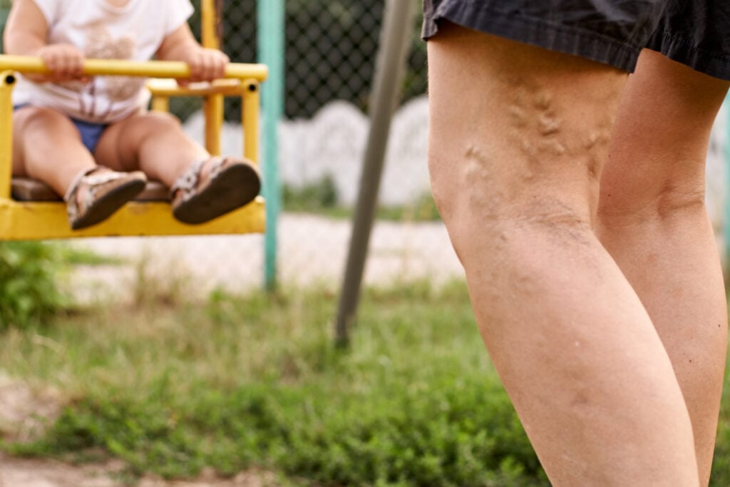
Varicose veins is a common condition, characterised by enlarged, swollen and twisted superficial veins, often a result of weakened or damaged valves inside the veins. Veins have one-way valves inside them that open and close to keep blood flowing toward the heart. Weak or damaged valves or walls in the veins can cause blood to pool and flow backward towards the ankle. This is called reflux and venous insufficiency. The calf muscles play a crucial role in pumping blood back to the heart, and when these valves malfunction, the calf muscles cannot effectively perform this function, contributing to varicose veins and swelling from the ankle up. While these highly visible vessels are the most common, it is possible to have varicose veins that are not visible on the skin surface.
Why we do this scan
The purpose of this scan is to assess for varicose veins and find where they are coming from. An ultrasound can examine the direction of the blood, the vein size and the pattern of venous insufficiency. This information assists the medical profession in determining if treatment is necessary and the treatment options available for your best care. Our ultrasound will also look to assess if there are any other identifiers contributing to your complaint.
Why choose us
The pattern of varicose veins in the legs is highly variable and specific to each patient. It takes years of specialised training to gain the skill of mapping out these veins so that nothing is missed. With our sonographer having >15 years of vein mapping experience and >10 years experience in the surgical procedures of varicose veins with multiple Surgeons, we provided accuracy and precision. Our technical drawings are meticulous to assist in any procedure you decide, eliminating the potential need for re-scans.
Risk factors
Risk factors for varicose veins include a family history, being female, pregnancy, obesity, prolonged standing or sitting and smoking, with age also playing a significant role. If you have one parent with varicose veins, your risk of developing them goes up by 40 percent, but if both your parents have them, your risk goes all the way up to 90 percent. Women are more likely to develop varicose veins than men, possibly due to hormonal changes during puberty, pregnancy, and menopause. Fluctuations in estrogen levels, such as during pregnancy or when taking birth control pills or hormone replacement therapy, can increase the risk. During pregnancy, increased blood volume and hormonal changes can put extra strain on the veins in the legs, increasing the risk of varicose veins. Being overweight and jobs or activities that require prolonged standing or sitting can increase pressure in the veins and contribute to varicose vein development. Smoking can damage the walls of blood vessels, making them more prone to becoming varicose.
Symptoms
Varicose veins can cause symptoms like aching, heaviness, itching or burning around the veins, and swelling in the legs, ankles, and feet, as well as skin discoloration. There can be cramps in the calves or thighs, often occurring at night and there may be a persistent urge to move the legs especially at night. Varicose veins may bleed more easily than other veins and in severe cases, they can lead to skin sores or ulcers that are slow to heal.
Complications
Most people don’t have complications from varicose veins. But in some people, an untreated varicose vein can cause ulcers, bleeding, inflammation or skin discoloration. While varicose veins themselves don't directly cause blood clots, they can increase the risk of developing a blood clot in the deep or superficial veins. Although varicose veins aren’t usually dangerous, you should visit your healthcare provider for an exam. If you’re concerned about how varicose veins look, or if they’re uncomfortable, treatments can help. You should see your provider as soon as possible if the skin or veins are experiencing bleeding, pain, redness, warmth to the touch, discolouration or swelling.
Deep Vs Superficial veins
The deep veins in the legs are located beneath the muscles and along bones, holding about 90% of the blood and carrying it back to the heart, while superficial veins are closer to the skin's surface and drain blood from the outer tissues into the deeper veins. Deep veins are crucial for returning blood to the heart while superficial veins are closer to the skin's surface and primarily drain blood from the skin and outer tissues, ultimately feeding into the deep veins. So if these superficial veins become “incompetant” (affected by venous reflux) they can be safely removed and the body does not miss them.
Spider veins
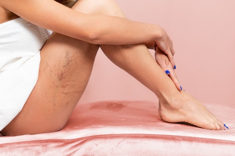
Spider veins, also known as telangiectasias, are small, damaged blood vessels visible on the skin, often appearing as thin, red, blue, or purple lines. They often look like spider webs or tree branches and are commonly found on the legs. Spider veins are typically harmless and don't cause any symptoms, but some people may experience mild discomfort or a burning sensation. Factors that increase the risk of developing spider veins include genetics, age, pregnancy, obesity, prolonged standing or sitting, and certain medications. Many people who have spider veins also have varicose veins and this is because they share many of the same causes and risk factors. Spider veins are not visible on ultrasound due to their size but prior to treatment, the ultrasound scan with determine if you also have varicose veins which may also need treatment for effective relief.
Preparation for the ultrasound
There is no patient preparation needed for this scan which will take up to 30 minutes for each leg. The scan assesses the entire leg from the groin crease to the ankle. We will require you to remove your shoes, socks and trousers/shorts. Very loose shorts, dresses and skirts don't need to be removed and can be lifted out of the way. We will ask for you to sit on the edge of our examination bed which will be lifted so your leg can comfortably rest over the edge as well as in a standing position on the ground. The procedure is usually painless and involves the sonographer applying a gel to your skin and moving the transducer over the area. We will need to periodically squeeze the calf and thigh to assess the blood flow.

CoreWave Vascular Ultrasound
Locations:
Suite 3, 46 Buckingham Drive
Wangara, WA 6065
Waikiki Specialist Centre
217 Willmott Drive
Waikiki, WA 6169
Duchess Medical Practice
4/69 Duchess Street
Busselton, WA 6280
More locations to come!
Contact Information:
Email: office@corewave.com.au
Phone: 0437 507 282
Business hours:
8am - 4pm
Monday to Friday
Monthly Saturdays
Fax: (08) 7947 7282